Home
/ Leg Bone Diagram Labeled : Pin On Good To Know _ An atlas of cat anatomy.
Leg Bone Diagram Labeled : Pin On Good To Know _ An atlas of cat anatomy.
Leg Bone Diagram Labeled : Pin On Good To Know _ An atlas of cat anatomy.. The lower leg is comprised of two bones the tibia and the smaller fibula. An atlas of cat anatomy. The knee joint is the largest joint in the body and is primarily a hinge joint, although some sliding and rotation. 15 photos of the leg bones anatomy diagram. See more ideas about muscle anatomy, human anatomy and physiology, body anatomy.
Below given knee diagram will help you to understand. I am not an expert at anatomy. 15 photos of the leg bones anatomy diagram. The upper leg is often called the thigh. Learn with flashcards, games, and more — for free.
Shin Splints Vector Illustration Leg Muscle Sport Trauma And Bone Pain Shin Splints Vector Illustration Leg Muscle Sport Canstock from comps.canstockphoto.com Leg bone anatomy diagram diagram of human leg human anatomy human leg bones anatomy stock photo download image now anatomy of the knee central coast orthopedic medical group A labeled diagram of the knee with an insight into its working. The outer and thinner bone of the two bones between the knee and ankle. Its lower end helps create the knee joint. Long bone femur label 12 photos of the long bone femur label , bone. Human anatomy bone diagram 12 photos of the human anatomy bone diagram human anatomy bone chart, human anatomy bone diagram, human anatomy diagram of bones, human anatomy skeleton diagram quiz. This is literally where the leg bone is connected to the hip bone. At the same time, the bones and joints of the leg and foot must be strong enough to support the body.
Labeled diagram of the human leg by xkeren on deviantart.
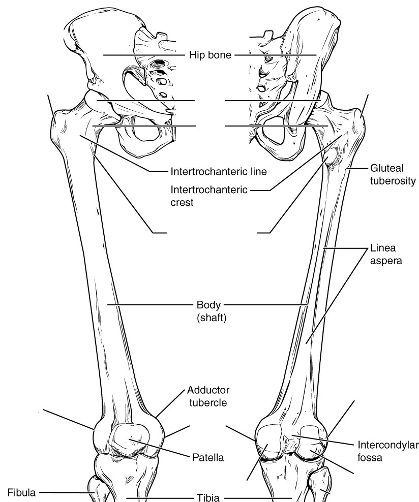
Labeled human leg bones created for use in leg bone. Our goal is that these leg anatomy worksheets pictures gallery can be a direction for you, bring you more references and also make you have a great day. If you are at standing and you move the leg around you can bring mobility to the femur in the acetabulum. Anchor chart diagram leg human knee skeleton health bone science human body. Labeled diagram of the human leg by xkeren on deviantart. Learn with flashcards, games, and more — for free. The acetabulum is where the head of the femur (top of the leg bone) goes. Bones of the leg and foot. Numbered one through five the bone that sits behind the big toe is no. This diagram depicts human leg bone anatomy.human anatomy diagrams show internal organs, cells, systems, conditions, symptoms and sickness information and/or tips for healthy living. Beside that, we also come with more related ideas as follows free printable human anatomy coloring pages, lower leg muscle diagram blank and lower limb bones unlabeled. The lower leg is comprised of two bones the tibia and the smaller fibula. Also called the thigh bone, this is the longest bone in the body.it.
The pubis, ischium, and ilium together constitute the pelvis while the thigh bone is the femur. The anatomy of the leg and foot bones. The femur, or thighbone, is the longest and largest bone in the human body. There is a quick overview of the bony structure of the pelvis. Our goal is that these leg anatomy worksheets pictures gallery can be a direction for you, bring you more references and also make you have a great day.
Lower Leg Bone Anatomy Anatomy Drawing Diagram from www.anatomylibrary99.com A heavy, long bone that forms the leg above the knee. The lower leg is comprised of two bones the tibia and the smaller fibula. Posted on june 4, 2014 by admin. It's the area that runs from the hip to the knee in each leg. The knee joint, you need a perfectly labeled diagram of the knee. This will help you to understand the mechanism as well as the working. The anatomy of the leg and foot bones. Normally, a smooth cushion of shiny white hyaline (or articular) cartilage about 1/4 inch thick covers the femoral head and the acetabulum.the articular cartilage is kept slick by fluid made in the synovial membrane (joint lining).
Related posts of diagram of leg bones long bone femur label.
Long bone femur label 12 photos of the long bone femur label , bone. Human anatomy diagrams show internal organs, cells, systems, conditions, symptoms and sickness information and/or tips for healthy living. This allows weight to be distributed either anteriorly or posteriorly throughout the foot. Terms in this set (7) femur. At the same time, the bones and joints of the leg and foot must be strong enough to support the body. This diagram depicts diagram leg bones anatomy. The anatomy of the leg and foot bones. The upper leg is often called the thigh. The acetabulum is where the head of the femur (top of the leg bone) goes. The bones of the leg and foot form part of the appendicular skeleton that supports the many muscles of the lower limbs. The knee joint is the largest joint in the body and is primarily a hinge joint, although some sliding and rotation. The bones together make up the hip. The talocrual joint is made up of three main bones.
Also called the thigh bone, this is the longest bone in the body.it. Normally, a smooth cushion of shiny white hyaline (or articular) cartilage about 1/4 inch thick covers the femoral head and the acetabulum.the articular cartilage is kept slick by fluid made in the synovial membrane (joint lining). To understand one of the most complex joints of our body i.e. Heart coloring pages free coloring pages anatomy coloring book coloring books coloring sheets science lessons life science science experiments apologia anatomy. The bones of the hip include the femur, the ilium, the ischium, and the pubis.
Pre Lab 2 Human Anatomy Lab Manual from uta.pressbooks.pub Related posts of diagram of leg bones long bone femur label. The bones of the leg are the femur, tibia, fibula and patella.the foot bones shown in this diagram are the talus, navicular, cuneiform, cuboid, metatarsals and calcaneus. Human anatomy diagrams show internal organs, cells, systems, conditions, symptoms and sickness information and/or tips for healthy living. The knee joint is the largest joint in the body and is primarily a hinge joint, although some sliding and rotation. Posted on june 4, 2014 by admin. The bones of the hip include the femur, the ilium, the ischium, and the pubis. The talocrual joint is made up of three main bones. The human leg, in the general word sense, is the entire lower limb of the human body, including the foot, thigh and even the hip or gluteal region.
Leg bone diagram labeled / lower limb bones (thigh, leg and foot) labeling page / click now to learn more about the bones, muscles, and soft tissues of these regions at kenhub!.
Labeled diagram of the human leg by xkeren on deviantart. This diagram depicts human leg bone anatomy.human anatomy diagrams show internal organs, cells, systems, conditions, symptoms and sickness information and/or tips for healthy living. Its lower end helps create the knee joint. At the same time, the bones and joints of the leg and foot must be strong enough to support the body. To understand one of the most complex joints of our body i.e. Ankle bones anatomy, arm bones anatomy, fibula anatomy, fibula fracture, hip bones anatomy, leg bones human body, foot, ankle bones anatomy, arm bones anatomy, fibula anatomy, fibula fracture, hip bones anatomy, leg bones human body. Normally, a smooth cushion of shiny white hyaline (or articular) cartilage about 1/4 inch thick covers the femoral head and the acetabulum.the articular cartilage is kept slick by fluid made in the synovial membrane (joint lining). The bones of the leg are the femur, tibia, fibula and patella.the foot bones shown in this diagram are the talus, navicular, cuneiform, cuboid, metatarsals and calcaneus. A heavy, long bone that forms the leg above the knee. I am not an expert at anatomy. This diagram of a feline skeleton shows you where all of your cat's bones are. Long bone femur label 12 photos of the long bone femur label , bone. Beside that, we also come with more related ideas as follows free printable human anatomy coloring pages, lower leg muscle diagram blank and lower limb bones unlabeled.
Labeled diagram of the human leg by xkeren on deviantart leg bone diagram. The bones of the hip include the femur, the ilium, the ischium, and the pubis.

